Multi-omics Platform
Cryo-EM Operation Team
Activities
Team Director
Shun-ichi Sekine
Cryo-electron microscopy (cryo-EM) is a powerful technique to analyze three-dimensional structure of biological macromolecules preserved in vitreous ice at cryogenic temperatures. Recent technological advances have dramatically improved the data quality, enabling high-resolution analysis of near-native structure of biomolecules. Consequently, cryo-EM is currently one of the primary methodologies for structural biology studies. The number of the cryo-EM structures deposited in the Electron Microscopy Data Bank and the Protein Data Bank has rapidly increased in recent years.
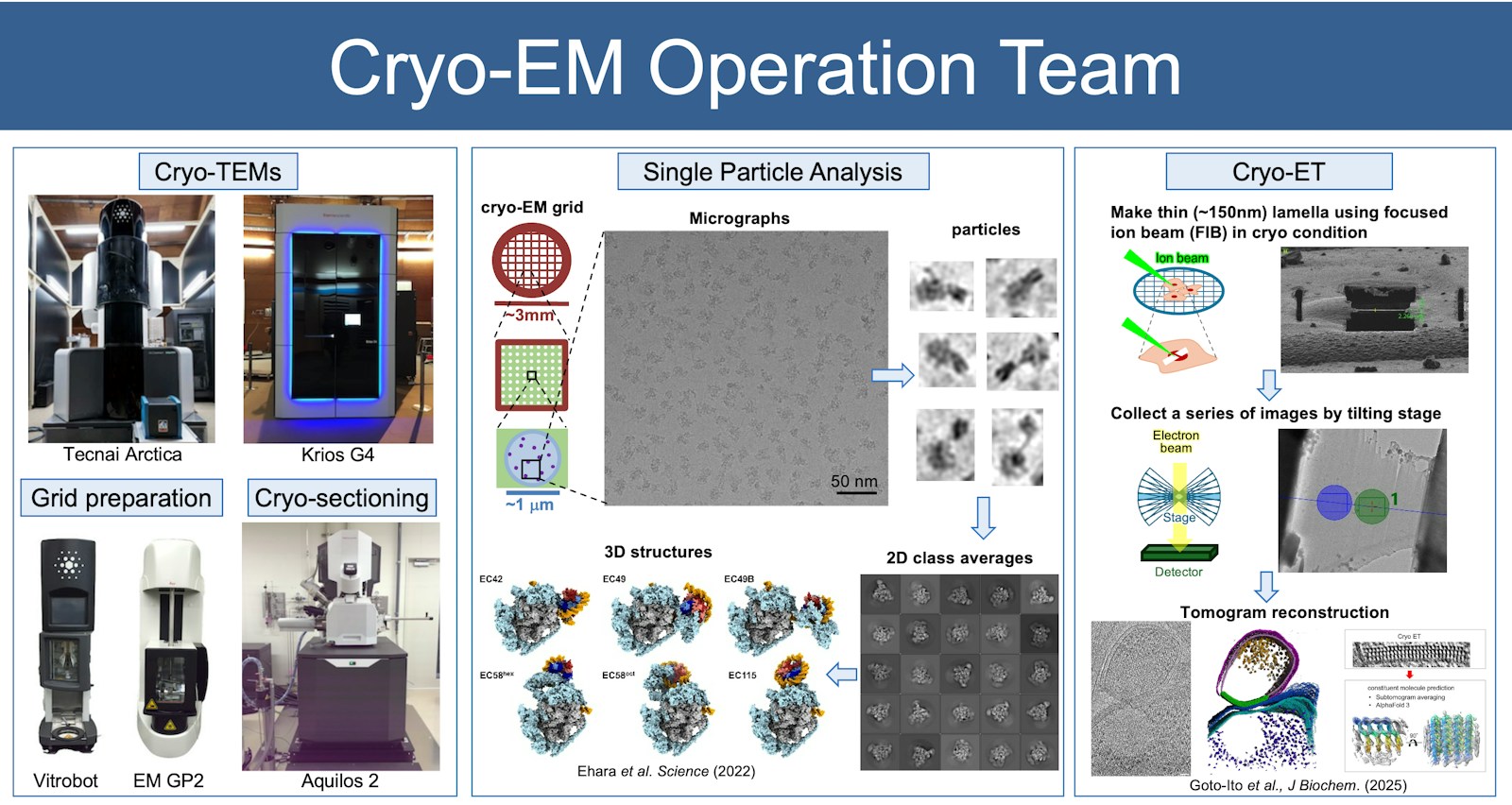
In RIKEN Yokohama, we operate two cryo-transmission electron microscopes (cryo-TEMs). Krios G4, a 300 kV cryo-TEM equipped with K3 and Falcon 4 direct detectors and an automated sample exchanger, serves as our main machine for the high-resolution cryo-EM data collection. Tecnai Arctica, a 200 kV cryo-TEM equipped with a K2 detector, is used for sample screening and routine data collection. For the cryo-grid preparation, we maintain automated plunge freezing systems for reproducible vitrification. To support cryo-electron tomography (cryo-ET) and MicroED, we have Aquilos2 cryo-FIB/SEM equipped with iFLM. These instruments and systems are available for shared use (https://cryoem.yokohama.riken.jp).
The Cryo-EM Operation Team is responsible for maintaining and operating these instruments to support a broad range of research activities. Through the shared use of cryo-EM, we contribute to the advancement of science and technology from academia to industry, while providing researchers with valuable training opportunities. While we support data collection for single particle analysis, we actively pursue technology development, with a particular emphasis on cryo-ET. Our goal is to establish robust cryo-ET workflows for in situ structural analysis of molecular entities within cells, tissues, and organs.
RIKEN Yokohama Cryo-EM Facility and available technology