Annual Report 2025
Credit to Cover Images
Acknowledgment and scientific context for the cover imagery featured in this report.
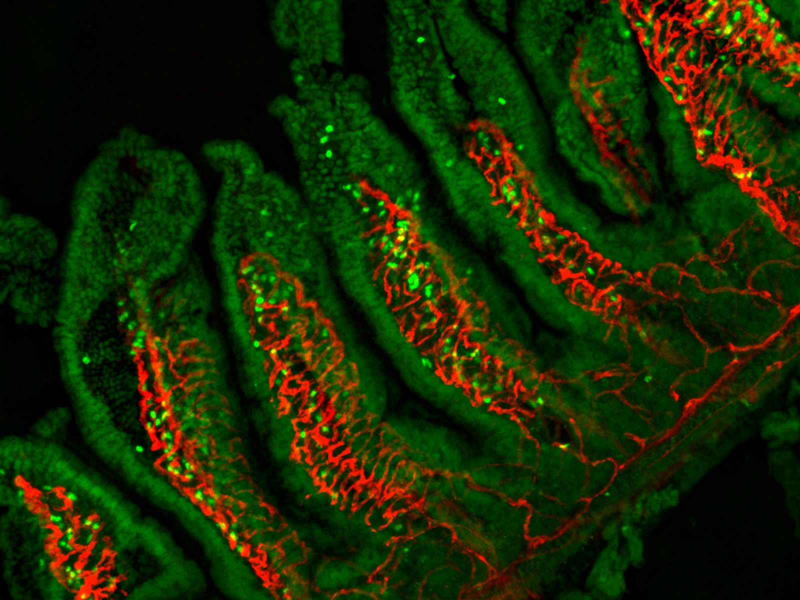
Fluorescence microscopy image of mouse intestinal villi showing epithelial cells (green) and blood vessels (red). This image demonstrates the intricate architecture of the intestinal mucosa, a key site for immune cell development and function.
Epithelial marker (green), Vascular marker (red)
Laboratory of Innate Immune Systems/ Laboratory of Immune Cell systems