Lab Activities
Laboratory for In situ Structural Biology
Research Activities
Team Director
Reiya Taniguchi
Cryo-electron tomography (cryo-ET) is an advanced imaging technique that combines transmission electron microscopy with computational three-dimensional reconstruction to visualize the three-dimensional architecture of frozen-hydrated biological specimens at sub-nanometer resolution. Recent methodological advances in cryo-ET have enabled us to visualize the intracellular environment in unprecedented detail and to analyze macromolecular complexes in situ at high resolution. The overarching aim of our research group is to fully exploit the potential of cryo-ET, to determine protein structures within their native cellular context, and to gain mechanistic insights into cellular phenomena from a structural perspective. Our current research focuses on two major questions: (i) the molecular basis of nuclear segmentation during cell differentiation and (ii) the regulatory mechanisms governing vesicle trafficking and cargo sorting at the Golgi apparatus.
As a newly established research group, our primary objective during this fiscal year was to set up and optimize a complete cryo-ET workflow, including sample freezing, sample thinning, data acquisition, and downstream data analysis. Using mammalian suspension cells as a model, we successfully established a pipeline for plunge-freezing cells, automated cryo-focused ion beam milling, and cryo-ET data acquisition. Using the acquired datasets, we performed template matching analysis and demonstrated reliable detection of multiple macromolecular assemblies, such as ribosomes, microtubules, and COPI coats, confirming the utility of the established workflow for future structure-based analyses.
Building on this foundation, we are currently performing cryo-ET data acquisition at the nuclear periphery and conducting systematic analyses to investigate nuclear morphology and chromatin organization. In parallel, we are developing a correlative cryo-light microscopy–based strategy to selectively target specific organelles for cryo-ET analysis. Through further optimization of these approaches, we aim to address the biological questions outlined above in the coming fiscal year.
Workflow for cryo-ET and in situ structural analysis
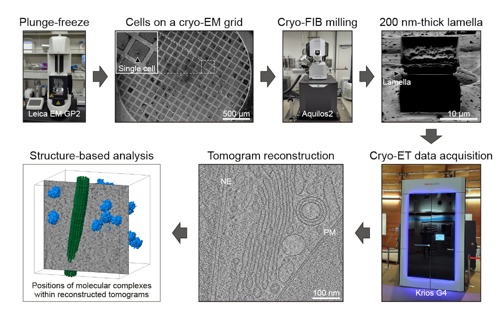
Overview of the typical workflow for cryo-ET sample preparation, data acquisition, and data processing. All data shown in this figure were acquired using instruments at the cryo-electron microscopy facility of IMS.
Invited Presentations
Taniguchi R, In situ cryo-ET reveals functional significance of the structural integrity of the NPC. The 98th annual meeting of the Japanese Biochemical Society, Kyoto, Japan, November 3rd-5th (2025)
Taniguchi R. Let’s start cryo-ET: Reality and pitfalls of the workflow. The 48th Annual Meeting of the Molecular Biology Society of Japan, Yokohama, Japan, December 3rd-5th (2025)
Taniguchi R. In situ cryo-ET uncovers the structural heterogeneity of the nuclear pore complex. The 81st Annual Meeting of the Japanese Society of Microscopy, Fukuoka, Japan, June 9th-11th (2025)