Lab Activities
Laboratory for Cancer Invasion and Metastasis
Research Activities

Team Director
Mikako Shirouzu
The major research interest of our laboratory is to search for target molecules that may be used for the treatment of intractable cancers, such as glioblastoma. Through RNA-sequencing analyses of genes encoding transmembrane proteins in glioma-initiating cells (GICs), we have identified herpes virus entry mediator (HVEM; also known as tumor necrosis factor receptor superfamily member 14 or TNFRSF14) as a potential target for treatment of glioblastoma. Among the subtypes of glioblastoma, we have found that HVEM is highly expressed in the mesenchymal subtype but not in non-mesenchymal subtypes of human glioblastoma cells and tissues. Silencing of HVEM expression resulted in decreased tumorigenic ability and prolonged survival of mice after intracranial injection of the GIC cells. We have identified major ligands, including APRIL (also known as tumor necrosis factor superfamily member 13 or TNFSF13), which are expressed in the mesenchymal subtype of GICs.
We have generated anti-human HVEM-VHH antibodies by immunizing alpaca. One of the anti-human HVEM-VHH antibodies was humanized and the Fc portion of human IgG was attached to obtain a longer half-life of the antibody in blood circulation. We have succeeded in three-dimensional analysis of the HVEM-anti-human HVEM-VHH antibody complex. Pre-clinical studies of the anti-human HVEM-VHH antibody, including pharmacokinetic studies and safety studies, have been performed. Generation of a master cell bank (MCB) stably producing HVR81 has been successfully conducted. Analyses of the antibody protein produced by the MCB cells have also been conducted.
In addition to HVR81, we have started to generate some other VHH antibodies by immunizing alpaca in collaboration with the School of Veterinary Medicine, Hokkaido University. We hope that such antibodies are useful for treatment of glioblastoma as well as some other cancers.
IgG Fc-fusion to extend half-life and humanization to diminish antigenicity of VHH.
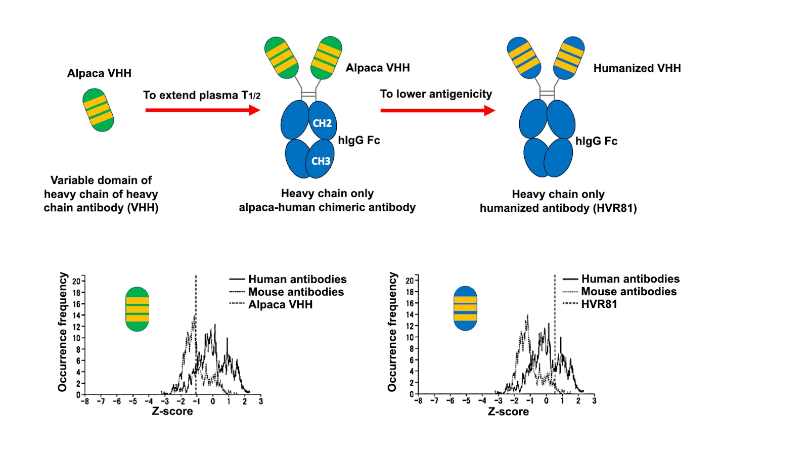
Conventional antibodies consist of two heavy chains and two light chains linked by disulfide bonds, whereas alpaca antibodies are composed solely of heavy chains, and their variable domains (known as VHHs or nanobodies) (upper left). Since the half-life of VHH in blood circulation is less than 12 hours, a technique, called chimerization, was employed to extend its half-life by fusing the VHH to IgG Fc of a different species including human while maintaining the original specificity and affinity (upper middle). Humanization of the VHH includes the grafting of amino acids in the complementarity-determining regions (CDRs) but also other framework amino acids critical for single chain formation and CDR orientation (upper right). The humanized antibody showed comparable binding activity to the original VHH with less antigenicity in human.
The degree of humanization was determined for sequences of the VHH (lower left) and the humanized VHH (HVR81) (lower right) in accordance with the integrated antibody sequence and structure analysis program (version 3.4.1) provided by Ebisu Co.